QPET
Quantitative PET
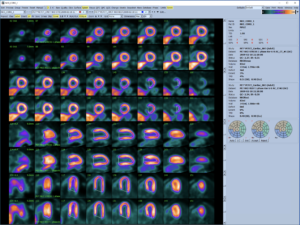
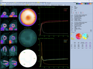
QPET provides cardiac function, perfusion, and Viability quantitation using gated and ungated MPI datasets: ED and ES volumes, ejection fraction, perfusion measures such as SSS/SRS/SDS and TPD, and Myocardial blood flow.
QPET: QGS+QPS with PET enhancements
An interactive stand-alone application for the automatic segmentation, quantification, and analysis of static, gated, and dynamic myocardial perfusion PET datasets.
Quantitative PET
An interactive stand-alone application for the automatic segmentation, quantification and analysis of static and gated myocardial perfusion PET, with support for both short axis and transverse datasets. It utilizes and integrates all of the features available in QGS and QPS.
PET Perfusion Databases created from scans of low-likelihood patients enables normal-limit based quantification of Rb-82 and N-13-ammonia perfusion PET images. Segmental stress and rest perfusion scores can be obtained using the familiar 17- or 20- segment models. Attenuation corrected PET/CT Rb-82 and N-13-ammonia normal limits are provided with QPET. These normal PET/CT databases have separate stress and rest files and combine data from both genders presenting no differences after attenuation correction.
The QPS and QGS contour algorithms have been optimized specifically for transverse PET data, providing a more reliable approach to identifying the heart and valve plane. This also includes an enhanced manual mode allowing reorienting of the transverse data. Once transverse data has been processed, it can be instantly reoriented to the standard short axis views associated with perfusion imaging for consistency in interpreting results.
Viability Quantification assess "hibernating myocardium" by calculation of relative regional changes between perfusion and viability in areas hypo-perfused on rest. Scar and mismatch parameters are reported as a percentage of the left ventricle. Extent and severity of scar and mismatch are displayed in polar map coordinates or as a 3D surface display. The program allows automatic scoring of scar using the 17- or 20-segment model. Simultaneous display of stress, rest and viability quantification results is possible. Stress images are not required though for the quantification of scar and mismatch.
Myocardial blood flow (MBF) and flow reserve (MFR) can be calculated from dynamic studies. The application allows for manual motion correction, with an upcoming update delivering automated motion correction for improved ease of use and throughput.
Request A Quote
Interested in purchasing the Cedars-Sinai Cardiac Suite?
Fill out a Quote Request
Call +1-844-CSMC-AIM
(844-276-2246), press 1 for Sales
Email sales@csaim.com


